Home
/ Diagram Of Major Muscles In Human Body - Basic Muscles Of The Body 10 Major Muscles Of The Human Body The Basic Muscles In The Human Human Muscular System Human Body Muscles Muscle Diagram / Outer clavicle, spine & acromion of scapula action:
Diagram Of Major Muscles In Human Body - Basic Muscles Of The Body 10 Major Muscles Of The Human Body The Basic Muscles In The Human Human Muscular System Human Body Muscles Muscle Diagram / Outer clavicle, spine & acromion of scapula action:
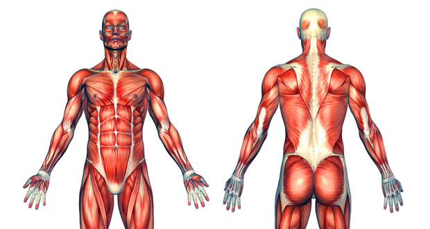
Diagram Of Major Muscles In Human Body - Basic Muscles Of The Body 10 Major Muscles Of The Human Body The Basic Muscles In The Human Human Muscular System Human Body Muscles Muscle Diagram / Outer clavicle, spine & acromion of scapula action:. In this image, you will find frontalis, orbicularis oculi, zygomaticus, masseter, orbicularis oris, sternocleidomasteoid, deltoid, pectoralis major, biceps brachii, iliopsoas, adductor longus, gastrocnemius. Physiology identification of muscles on the human body. Lower thoracic, lumbar vertebrae and sacrum: As seen in the image below, a muscle is arranged in a basic pattern of bundled fibers separated by protective layers. The human body consists of many muscles.
This is what happens in the body. Human body muscle system, the muscles of the human body that work the skeletal system, that are under voluntary control, and that are concerned with movement, posture, and balance. If someone wants a healthy and good life, one must understand his body. Posted on june 7, 2016 by admin. Pectoralis minor small anterior, upper chest deep to pec major
Human Muscles Major Muscles Structure Fibre Types Teachpe Com from www.teachpe.com All the major muscle groups of the body from front and back. This type of muscle creates movement in the body. The muscles of the trunk human anatomy and physiology lab bsb 141. Physiological diagram of the muscles, joints and animal mechanics of the human body date: The abdominal muscles are shown in red, it is very easy to see from this diagram how a six pack is made, and also why some people have an eight pack. Human body muscles human muscle anatomy human anatomy and physiology major muscles human anatomy picture muscular system anatomy muscle diagram anatomy bones anatomy organs. They cause motion and produce a force that the body uses to move and manipulate the body. Click on the name of the muscle, or the image, to see weight training exercises.
In this image, you will find frontalis, orbicularis oculi, zygomaticus, masseter, orbicularis oris, sternocleidomasteoid, deltoid, pectoralis major, biceps brachii, iliopsoas, adductor longus, gastrocnemius.
1870 the muscles of the human body. If someone wants a healthy and good life, one must understand his body. When you are taking anatomy and physiology you will be required to identify major muscles in the human body diagram of muscles in the body. The four huge muscles are namely rectus femoris , vastus lateral , vastus intermedius and vastus medialis. Muscular system muscles of the human body muscle diagrams of major muscles exercised in weight training meet some muscles science learning hub a fully labelled human body muscle diagram fit and healthy map of body muscles tendernessco muscles the human body anatomy physiology. The best way to strengthen back muscles is in a static position. Each of these muscles is a discrete organ constructed of skeletal muscle tissue, blood vessels, tendons, and nerves. Shoulder girdle muscles trapezius flat sheet of muscle on upper back. Physiological diagram of the muscles, joints and animal mechanics of the human body date: Brings shoulders and arms back to body. Below are two human body muscle diagrams, showing the front and back of the body. The muscles of the trunk human anatomy and physiology lab bsb 141. Diagram of muscle in the body wiring diagram database.
This type of muscle creates movement in the body. Related to the function of movement is the muscular system's second function: The skeletal muscles are continually making fine adjustments that hold the body in stationary positions. Skeletal muscle, which exists throughout the body the body's collective muscle tissue constitutes its muscle mass. Located immediately below the skin) muscles of the body.
Learn All Muscles With Quizzes And Labeled Diagrams Kenhub from thumbor.kenhub.com Pectoralis minor small anterior, upper chest deep to pec major Physiological diagram of the muscles, joints and animal mechanics of the human body date: Posted on june 7, 2016 by admin. Muscle diagrams are a great way to get an overview of all of the muscles within a body region. It is located in the upper front part of the leg. This type of muscle creates movement in the body. The pectoralis major originates along the clavicle down the sternum and across the ribs and inserts into the humerus. Elevation, depression, adduction & up.
This is what happens in the body.
Lower thoracic, lumbar vertebrae and sacrum: Nevertheless, the exact number is difficult to define. Human body muscle system, the muscles of the human body that work the skeletal system, that are under voluntary control, and that are concerned with movement, posture, and balance. Major body muscles and diagrams » body muscles and diagrams following labelled diagram of human anterior muscles includes some required by itec diploma in all. When you are taking anatomy and physiology you will be required to identify major muscles in the human body diagram of muscles in the body. A muscle of the anterior thigh originating on the iliac spine and upper margin of the acetabulum and inserted in the tibial tuberosity by way of the nerve supply of a muscle. A chest muscle that pulls the arm in towards the body. Extensor muscles of the hand. Skeletal muscle, which exists throughout the body the body's collective muscle tissue constitutes its muscle mass. Human body muscle system, the muscles of the human body that work the skeletal system, that are under voluntary control, and that are concerned with movement, posture, and balance. The skeletal muscles are continually making fine adjustments that hold the body in stationary positions. Muscles graph diagram page 6. This is one of the internal rotator muscles that attach the humerus and internally rotate the arm.
This diagram depicts picture of female reproductive system diagram 1024×1204 with parts and labels. The abdominal muscles are shown in red, it is very easy to see from this diagram how a six pack is made, and also why some people have an eight pack. In this image, you will find frontalis, orbicularis oculi, zygomaticus, masseter, orbicularis oris, sternocleidomasteoid, deltoid, pectoralis major, biceps brachii, iliopsoas, adductor longus, gastrocnemius. The pectoralis major originates along the clavicle down the sternum and across the ribs and inserts into the humerus. The skeletal muscles are continually making fine adjustments that hold the body in stationary positions.
Diagram Of Major Muscles In Human Body Graph Diagram from graphdiagram.com Diagram of muscle in the body wiring diagram database. Located immediately below the skin) muscles of the body. The skeletal muscles are continually making fine adjustments that hold the body in stationary positions. Skeletal muscle, which exists throughout the body the body's collective muscle tissue constitutes its muscle mass. Brings shoulders and arms back to body. Moves humerus (arm) to chest. The abdominal muscles are shown in red, it is very easy to see from this diagram how a six pack is made, and also why some people have an eight pack. Major muscles on the front of the body yoga pain management.
The abdominal muscles are shown in red, it is very easy to see from this diagram how a six pack is made, and also why some people have an eight pack.
Physiology identification of muscles on the human body. This diagram depicts picture of female reproductive system diagram 1024×1204 with parts and labels. Lower thoracic, lumbar vertebrae and sacrum: Below are two human body muscle diagrams, showing the front and back of the body. As seen in the image below, a muscle is arranged in a basic pattern of bundled fibers separated by protective layers. The human body consists of many muscles. In this image, you will find frontalis, orbicularis oculi, zygomaticus, masseter, orbicularis oris, sternocleidomasteoid, deltoid, pectoralis major, biceps brachii, iliopsoas, adductor longus, gastrocnemius. Almost every muscle constitutes one part of a pair of identical bilateral muscles, found on both sides, resulting in approximately 320 pairs of muscles, as presented in this article. The best way to strengthen back muscles is in a static position. When you are taking anatomy and physiology you will be required to identify major muscles in the human body diagram of muscles in the body. 1870 the muscles of the human body. Major muscles on the front of the body yoga pain management. Brings shoulders and arms back to body.
They cause motion and produce a force that the body uses to move and manipulate the body diagram of muscles in body. The muscles of the trunk human anatomy and physiology lab bsb 141.
